Translational Research Unit (TRU) Platform

The Team of the Translational Research unit (TRU). Please contact us at tru.igmp@unibe.ch

TRU Research Platform

Projects

The TRU supports research projects using patient-derived rest material and animal tissues from investigators on and off campus. We are involved in national (e.g. SAKK) and international collaboratives, where expertise in tissue handling and biomarkers is needed. More than 600 requests are processed per year. Services and associated pricing can be found here.

Biobanking

Our biobank handles more than 5’000 tissue samples per year and stores more than 37’000 aliquots of tissue. We work with Human Research Act compliant workflows, following SOPs documented in a Quality Management System and received the ISO accreditation for the interfaces with the clinical pathology lab (ISO 17025/15189).

Histology

Expertise in histology techniques is the foundation of our lab. We process tissues and make sections for many downstream applications such as conventional staining, immunohistochemistry and in situ hybridisation. We also support in clinical studies management (SAKK, Genfit NASH, etc)

Tissue visualisation

Our lab establishes a large number of antibodies per year in human and animal tissue. We validate all new antibodies (commercial and homemade) in positive and negative cell lines. Our catalog now exceeds 800 antibodies. We perform single and double immunostaining, single and double mRNA in situ hybridisation (RNAScope®), TUNEL, Multiplex immunofluorescence (OPAL and Ultivue) and set up stainings on chamber slides for cell cultures. We set up protocols for researchers and are always ready to experiment with new methods.

Slide scanning and access to digital scans

We offer slide scanning to researchers interested in archiving slides, performing digital image analysis, preparing next-generation Tissue Microarrays (ngTMA®), or wishing to publish high-quality images. Scans can be uploaded to an Image Management Software accessible to external collaborators. We use one of three in-house scanners for this purpose.

Digital Image Analysis (DIA)

TRU performs DIA on whole slide images and tissue microarrays using in-house tools (Scorenado), open source software (QuPath) and Indica labs Halo. Examples include tumor-lymphocyte proximity studies, quantification of breast cancer biomarker panels, and annotation of whole slide images or tissue microarray spots. Our computational pathology team uses machine-learning and deep learning to answer clinically relevant questions with direct implications for diagnostic routine.

Next-generation Tissue Microarrays

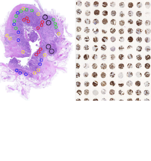
We construct tissue microarrays based on digitally annotated scans. This allows researchers to capture the most important regions within their tissues for their downstream analysis. We call our approach next-generation Tissue Microarrays (ngTMA®). Since 2012, our lab has constructed almost 1000 ngTMA blocks for studies related to precision medicine, tumor heterogeneity, rare diseases, animal models and establishment of new tissue visualization techniques (e.g. CODEX and OPAL). We have collected images from more than 1 million tissue spots, corresponding to several thousand patients with annotated clinicopathological data, which can now be explored using artificial intelligence.

COMPATH

Whatever we do can apply to animal tissues, from histology to visualisation, digital pathology, image analysis and ngTMA construction. Together with the Institute of Animal Pathology, we are delighted to collaborate where morphology and histopathology evaluation as well as technology is concerned.